Overview
he accessory navicular (os navicularum or os tibiale externum)is an extra bone or piece of cartilage located on the inner sideof the foot just above the arch. It is incorporated within the posterior tibial tendon, which attaches in this area. An accessory navicular is congenital (present at birth). It is not part of normal bone structure and therefore is not present in most people. What is Accessory Navicular Syndrome? People who have an accessory navicular often are unaware of the condition if it causes no problems. However, some people with this extra bone develop a painful condition known as accessory navicular syndrome when the bone and/or posterior tibial tendon are aggravated.

Causes
Like all painful conditions, ANS has a root cause. The cause could be the accessory navicular bone itself producing irritation from shoes or too much activity. Often, however, it is related to injury of one of the structures that attach to the navicular bone. Structures that attach to the navicular bone include abductor hallucis muscle, plantar calcaneonavicular ligament (spring ligament) parts of the deltoid ligament, posterior tibial tendon.
Symptoms
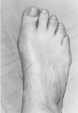
Many people have accessory (?extra?) naviculars (figure 1) - a prominent extra bone extending from the navicular bone. Most accessory naviculars are completely asymptomatic. However, some individuals will develop pain on the inside of their midfoot. Pain may occur from the pressure of the shoe ware against the prominence, irritating either the bone itself or the fibrous junction where the accessory bone meets the regular navicular. Alternatively, the fibrous junction or interface may become painful as a result of tension applied by the posterior tibial tendon through its connection or insertion at that site. Often, individuals will be asymptomatic for years, however, a new pair of shoes or a change in their activity level can cause symptoms. The accessory navicular itself typically develops during adolescence, when the two areas of the navicular bone fail to fuse together.
Diagnosis
Your podiatrist will most likely diagnose accessory navicular syndrome by making a visual study of the area, checking whether the shape of your foot and your ability to move it indicate there?s an accessory navicular lurking around. He or she may push on the prominence on your foot to check to see if it hurts, and may ask you to walk around in order to ascertain how your gait is affected. In order to get a certain diagnosis, your podiatrist will need some way to see the inside of your foot, which will most likely involve getting X-rays, or possibly an MRI or some other scan of your foot?s interior.
Non Surgical Treatment
Treating accessory navicular syndrome is focused on relieving symptoms. Some treatment methods are Icing to reduce swelling. Immobilization with a cast or walking boot to reduce inflammation and promote healing. Medications to reduce pain and inflammation. Physical therapy to strengthen muscles. Orthotics to support the arch. Surgery may be needed to remove the accessory bone and reshape the area if other methods are not successful.
Surgical Treatment
In my experience, the Modified Kidner procedure is one of the most reliable operations for reducing arch pain associated with an accessory navicular bone (a.k.a. os tibial externum). You can also use this procedure to treat a prominence at the inner aspect of the arch, which has been caused by an enlarged navicular bone. The most common patients to visit our office with these problems are between the ages of 8 and 15 and are involved in activities like ice skating, ballet and soccer.
he accessory navicular (os navicularum or os tibiale externum)is an extra bone or piece of cartilage located on the inner sideof the foot just above the arch. It is incorporated within the posterior tibial tendon, which attaches in this area. An accessory navicular is congenital (present at birth). It is not part of normal bone structure and therefore is not present in most people. What is Accessory Navicular Syndrome? People who have an accessory navicular often are unaware of the condition if it causes no problems. However, some people with this extra bone develop a painful condition known as accessory navicular syndrome when the bone and/or posterior tibial tendon are aggravated.
Causes
Like all painful conditions, ANS has a root cause. The cause could be the accessory navicular bone itself producing irritation from shoes or too much activity. Often, however, it is related to injury of one of the structures that attach to the navicular bone. Structures that attach to the navicular bone include abductor hallucis muscle, plantar calcaneonavicular ligament (spring ligament) parts of the deltoid ligament, posterior tibial tendon.
Symptoms
Many people have accessory (?extra?) naviculars (figure 1) - a prominent extra bone extending from the navicular bone. Most accessory naviculars are completely asymptomatic. However, some individuals will develop pain on the inside of their midfoot. Pain may occur from the pressure of the shoe ware against the prominence, irritating either the bone itself or the fibrous junction where the accessory bone meets the regular navicular. Alternatively, the fibrous junction or interface may become painful as a result of tension applied by the posterior tibial tendon through its connection or insertion at that site. Often, individuals will be asymptomatic for years, however, a new pair of shoes or a change in their activity level can cause symptoms. The accessory navicular itself typically develops during adolescence, when the two areas of the navicular bone fail to fuse together.
Diagnosis
Your podiatrist will most likely diagnose accessory navicular syndrome by making a visual study of the area, checking whether the shape of your foot and your ability to move it indicate there?s an accessory navicular lurking around. He or she may push on the prominence on your foot to check to see if it hurts, and may ask you to walk around in order to ascertain how your gait is affected. In order to get a certain diagnosis, your podiatrist will need some way to see the inside of your foot, which will most likely involve getting X-rays, or possibly an MRI or some other scan of your foot?s interior.
Non Surgical Treatment
Treating accessory navicular syndrome is focused on relieving symptoms. Some treatment methods are Icing to reduce swelling. Immobilization with a cast or walking boot to reduce inflammation and promote healing. Medications to reduce pain and inflammation. Physical therapy to strengthen muscles. Orthotics to support the arch. Surgery may be needed to remove the accessory bone and reshape the area if other methods are not successful.
Surgical Treatment
In my experience, the Modified Kidner procedure is one of the most reliable operations for reducing arch pain associated with an accessory navicular bone (a.k.a. os tibial externum). You can also use this procedure to treat a prominence at the inner aspect of the arch, which has been caused by an enlarged navicular bone. The most common patients to visit our office with these problems are between the ages of 8 and 15 and are involved in activities like ice skating, ballet and soccer.
